Description
Here's a detailed product description for Histopathology and Immunohistochemistry services, formatted in Markdown:
Comprehensive Histopathology & Immunohistochemistry Services: Precision Tissue Analysis for Research & Diagnostics
Unlock profound insights into disease mechanisms, therapeutic efficacy, and biomarker discovery with our state-of-the-art Histopathology and Immunohistochemistry (IHC) services. We offer a full spectrum of tissue analysis solutions, from meticulous sample preparation to expert pathological interpretation, designed to support your research, preclinical development, and diagnostic needs with unparalleled accuracy and efficiency.
Why Tissue Analysis Matters
Tissue analysis is the cornerstone of biological and medical research, offering irreplaceable spatial context and cellular resolution. Whether identifying subtle morphological changes, localizing specific proteins, or characterizing cellular populations, our services provide the critical data necessary for informed decision-making in diverse fields.
Our Core Services
1. Histopathology: The Foundation of Tissue Assessment
Histopathology provides the essential morphological context for understanding tissue health and disease. Our experienced pathologists and skilled technicians ensure the highest quality results from sample processing to microscopic evaluation.
Key Histopathology Services Include:
- Tissue Processing & Embedding:
- Fixation: Formalin fixation, cryopreservation, or specialized fixation methods tailored to your sample type and downstream analysis.
- Grossing: Macroscopic examination and precise trimming of tissues by expert personnel.
- Paraffin Embedding: Meticulous embedding of tissues in paraffin blocks for long-term storage and sectioning.
- Microtomy & Sectioning:
- Paraffin Sectioning: Production of high-quality, uniform tissue sections (typically 3-5 µm) from paraffin blocks.
- Frozen Sectioning (Cryosectioning): Rapid sectioning of fresh or frozen tissues for quick diagnostic evaluation or specific research applications (e.g., RNA/DNA extraction compatibility).
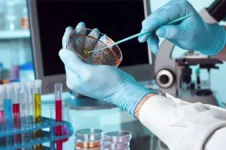
- Routine & Special Staining:
- Hematoxylin & Eosin (H&E): The gold standard for foundational morphological assessment, providing clear visualization of cellular and tissue architecture.
- Special Stains: A comprehensive panel of histochemical stains to highlight specific tissue components (e.g., collagen, elastic fibers, mucin, lipids, microorganisms) including:
- Masson's Trichrome, Picrosirius Red, Reticulin, PAS, Alcian Blue, Prussian Blue, Congo Red, Oil Red O, and many more.
- Expert Pathological Evaluation & Reporting:
- Board-certified veterinary and/or human pathologists provide detailed, quantitative, and qualitative assessments.
- Comprehensive reports with macroscopic and microscopic findings, severity scores, and descriptive pathology.
- Toxicopathology studies, efficacy studies, and diagnostic evaluations.
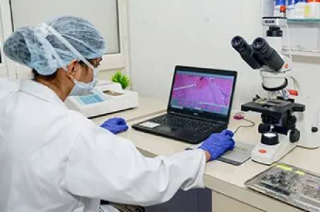
- Digital pathology solutions for remote review and quantitative image analysis.
2. Immunohistochemistry (IHC): Pinpointing Molecular Targets
Immunohistochemistry utilizes the specificity of antibody-antigen binding to visualize and localize specific proteins, antigens, or biomarkers directly within tissue sections. It is indispensable for confirming cell identity, assessing protein expression, and identifying disease targets.
Our Advanced IHC Capabilities:
- Primary Antibody Validation & Optimization:
- Extensive experience with thousands of commercially available antibodies.
- Custom development and optimization protocols for novel or difficult-to-detect targets.
- Chromogenic & Fluorescent IHC:
- Chromogenic Detection (e.g., DAB, AP): High-resolution visualization of single or dual markers. Ideal for morphological correlation and archiving.
- Fluorescent Detection: Multiplexing capabilities for simultaneous detection of 2-5+ markers on a single slide, enabling complex cellular interaction studies.
- Multiplex IHC & Immunofluorescence (mIHC/mIF):
- Simultaneously detect multiple protein targets within a single tissue section, providing spatial relationships and co-expression analysis.
- Advanced image acquisition and analysis for complex cellular phenotyping.
- Automated IHC Staining:
- High-throughput, reproducible, and standardized staining using automated platforms, minimizing variability and increasing efficiency.
- Quantitative Image Analysis:
- Advanced software solutions for objective quantification of IHC staining (e.g., positive cell counting, intensity scoring, H-score, tumor microenvironment analysis, segmentation).
- Detailed data output and statistical analysis support.
- Custom IHC Panel Development:
- Tailored panels for specific research questions, including biomarker discovery, companion diagnostic development, and target engagement studies.
- Specialized IHC Applications:
- In Situ Hybridization (ISH): For detection of DNA/RNA targets (e.g., mRNA, viral genomes).
- Immunofluorescence (IF) on Frozen/Fresh Tissue: Ideal for sensitive antigens or specific research applications where paraffin processing may alter epitopes.
Applications & Benefits
Our comprehensive services are ideal for a diverse range of applications, empowering clients in:
- Preclinical Drug Development: Efficacy studies, safety assessment, toxicology, target validation.
- Clinical Trials & Diagnostics: Biomarker identification, patient stratification, companion diagnostics, prognostic indicators.
- Biomarker Discovery & Validation: Identifying novel protein markers for disease progression, therapy response, or diagnosis.
- Academic & Basic Research: Understanding disease mechanisms, cellular biology, and tissue pathology.
- Toxicopathology: Identifying and characterizing tissue changes induced by compounds.
- Veterinary Pathology: Diagnostic support, research studies in animal models.
- Genetically Modified Organism (GMO) Characterization: Phenotyping and assessing morphological changes.
Why Partner With Us?
- Expert Pathologists: Access to a team of board-certified pathologists with specialized expertise in various therapeutic areas and animal models.
- State-of-the-Art Laboratory: Equipped with advanced instrumentation for high-throughput processing, staining, imaging, and quantitative analysis.
- Customized Solutions: Flexible service models and bespoke protocol development to meet your unique project requirements.
- Quality & Compliance: Adherence to rigorous quality control standards, including GLP-compliant studies where required, ensuring reliable and auditable data.
- Timely & Efficient Delivery: Streamlined workflows and dedicated project management to ensure rapid turnaround times without compromising quality.
- Comprehensive Reporting: Detailed, high-quality reports accompanied by high-resolution images, raw data, and expert consultation.
- Dedicated Project Management: A single point of contact to ensure seamless communication and project oversight.
Our Seamless Workflow
- Consultation & Project Design: Discuss your specific needs, experimental design, and desired outcomes with our scientific team.
- Sample Submission: Secure and trackable submission of your tissue samples (blocks, slides, or fresh tissue).
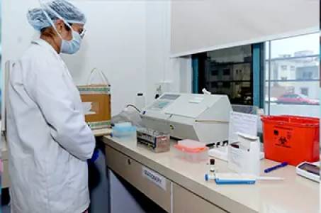
- Processing & Staining: Meticulous tissue processing, sectioning, and application of chosen H&E, special stains, or IHC panels.
- Pathological Evaluation & Image Acquisition: Expert microscopic analysis, scoring, and high-resolution digital imaging.
- Data Analysis & Reporting: Comprehensive reports, quantitative data, and expert interpretation.
- Delivery & Consultation: Secure delivery of results, accompanied by a follow-up consultation to discuss findings.
Get Started Today
Accelerate your research and enhance your diagnostic capabilities with our premium Histopathology and Immunohistochemistry services.
Contact us for a free consultation and a custom quote. Let our expertise become your advantage.
[Your Company Name] [Your Website] [Your Phone Number] [Your Email Address] [Your Location, if relevant]